Introduction
When you experience persistent back pain, neck discomfort, or suspect a spinal injury, your healthcare provider often recommends diagnostic imaging to determine the underlying cause. Whether you live in a major city, small town, or anywhere searching for quality spine imaging, X-rays of the spine, neck, or back remain one of the most commonly ordered, accessible, and cost-effective diagnostic tools in modern medicine. From clinics in Spain offering advanced diagnostics to healthcare facilities worldwide, this imaging technique helps millions of patients every year. Understanding how spinal X-rays work empowers you to make informed decisions about your healthcare. This comprehensive guide explores everything you need to know about X-ray imaging of the spine, including how the procedure works, what conditions can be detected, and how X-rays compare to other imaging modalities like MRI and CT scans.
What Are Spinal X-Rays?
X-rays of the spine, neck, or back are radiographic imaging techniques that use electromagnetic energy beams to produce two-dimensional images of the spine and surrounding structures. Whether you need a spine X-ray at a Barcelona clinic or imaging at any international facility, the technology remains consistent. Unlike more complex imaging methods, traditional X-rays capture clear, detailed images of bones and can reveal alignment issues, fractures, and degenerative changes within minutes. The procedure is quick, non-invasive, and requires minimal preparation, making it an ideal first-line diagnostic tool for many spinal conditions. The human spine consists of several regions, and doctors may request X-rays from specific areas depending on your symptoms:
Cervical spine
The upper portion consists of seven vertebrae that form your neck.
Thoracic spine: The middle section with twelve vertebrae attached to the rib cage
Lumbar spine: The lower back region contains five large vertebrae
Sacrum and coccyx: The base of the spine, where additional bones fuse
Understanding Soft Tissue Neck X-Ray Imaging
When patients experience neck pain, swelling, or symptoms suggestive of airway issues, physicians often order a soft tissue neck X-ray to evaluate structures beyond bone. A soft tissue neck X-ray is particularly valuable for assessing conditions affecting the soft tissues surrounding the cervical spine, including muscles, ligaments, and the airway. The soft tissue neck X-ray technique involves positioning the patient carefully to ensure proper visualization of anatomical structures. During this imaging procedure, patients typically stand or sit upright with the neck extended and chin tucked. The X-ray beam is directed horizontally at the C4 vertebra level, allowing radiologists to examine the prevertebral space, epiglottis, and various pharyngeal structures.
Clinical Applications of Soft Tissue Neck Imaging
A soft tissue neck X-ray can help physicians diagnose:
- Retropharyngeal abscesses from serious infections
- Foreign body obstructions in the airway
- Epiglottitis or croup conditions
- Prevertebral space widening indicates infection
- Cervical spine alignment abnormalities
- Loss of normal cervical lordosis
The X-ray of soft tissue neck provides critical information about whether soft tissue swelling or infection is compromising the airway, which requires immediate medical attention. Radiologists carefully measure the prevertebral soft tissue space, comparing it to adjacent vertebral bodies to identify abnormal widening that suggests serious infection or abscess formation. When evaluating X-rays of soft tissue neck findings, clinicians consider multiple factors to ensure comprehensive diagnosis and appropriate treatment planning.
Can You See a Herniated Disk on an X-Ray?
One of the most frequently asked questions by patients is: “Can you see a herniated disk on an X-ray?” The straightforward answer is no—a standard X-ray cannot directly visualize a herniated disk because herniated discs involve soft tissue displacement rather than bone pathology.
Understanding X-Ray Limitations for Disc Herniation
Can you see a herniated disk on an X-ray?
To understand why not, consider how X-rays work. X-ray imaging is specifically designed to capture images of dense structures like bones. Since herniated discs involve the displacement of the intervertebral disc (a soft tissue structure), they fall outside the diagnostic capabilities of conventional X-ray imaging. The disc material itself and the nerve compression it causes simply don’t appear on standard radiographs.
However, can you see a herniated disk on an X-ray? In an indirect way?
Yes. While X-rays cannot show the herniated disc itself, they can reveal secondary changes that suggest disc pathology:
- Narrowed disc space: The distance between vertebrae may appear reduced on X-ray
- Bone spurs: Degenerative changes called osteophytes may develop adjacent to the affected disc
Vertebral misalignment: The vertebrae may shift position if the disc has herniated significantly - Loss of normal spinal curve: Changes in the spine’s normal lordosis or kyphosis can indicate underlying disc problems
When Advanced Imaging Is Necessary
If your physician suspects disc herniation and needs a definitive diagnosis, they’ll typically recommend advanced imaging modalities rather than relying on x ray spain or CT scan facilities alone:
MRI (Magnetic Resonance Imaging)
The gold standard for soft tissue visualization, MRI provides detailed images of discs, nerves, and the spinal cord.
CT (Computed Tomography) scans
Offers superior bone detail and can visualize both bone and some soft tissue abnormalities
Myelography
Involves injecting contrast material to enhance the visualization of the spinal cord and nerve roots
Different Types of Spine X-Rays and Their Indications
Healthcare providers order different X-ray views depending on the clinical situation and region of the spine requiring evaluation. Each view reveals specific information about spinal anatomy and pathology, whether you’re at a major teaching hospital or a small clinic in Spain.
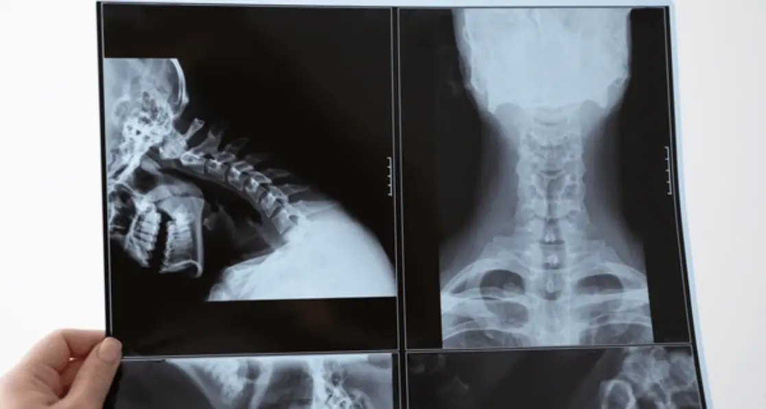
Cervical Spine X-Rays
- Anteroposterior (AP) View: Shows the front-to-back alignment of the cervical vertebrae (C3-C7), useful for detecting compression fractures and spinous process fractures
- Lateral View: Reveals the side profile of the cervical spine, helpful for diagnosing trauma, osteoarthritis, and spondylosis
- Oblique Views: Specifically designed to visualize the intervertebral foramina (nerve root passages) to detect stenosis or nerve compression
- Odontoid (Open Mouth AP) View: Focuses on the C1 and C2 vertebrae and is essential for ruling out fractures of the atlas or axis
- Flexion-Extension Views: Performed while the patient bends forward and backward to assess spinal stability and detect abnormal movement
Thoracic Spine X-Rays
AP and Lateral Views: Standard views for evaluating trauma, chronic diseases, compression fractures, and postoperative complications. These views help clinicians assess spinal alignment, detect fractures, monitor healing, and identify degenerative changes in the thoracic region.
Lumbar Spine X-Rays
AP/PA View: Shows the front-to-back or back-to-front alignment of lumbar vertebrae, useful for trauma and chronic disease assessment
- Lateral View: Provides a side view of the lumbar spine to evaluate alignment and disc space height
- Oblique Views: Specifically target the pars interarticularis to detect spondylolisthesis or pars defects
- Flexion-Extension Views: Help determine spinal stability by showing movement at each segment
What Conditions Can X-Rays Detect?
Spinal X-rays are effective at identifying numerous bone-related conditions affecting the spine, neck, or back. Common diagnoses made with X-ray imaging include:
Fractures and Traumatic Injuries
X-rays are excellent at detecting broken bones, including compression fractures, vertebral body fractures, spinous process fractures, and displaced vertebrae. In trauma situations, X-rays provide a rapid assessment of whether spinal injury has occurred, though CT imaging is increasingly preferred for more complex injuries. Whether you need evaluation at a facility offering spine X-ray services or elsewhere, prompt fracture detection is critical.
Degenerative Changes
Age-related wear and tear on the spine produces characteristic changes visible on X-rays:
- Bone spurs (osteophytes)
- Disc space narrowing
- Joint space narrowing in the facet joints
- Sclerosis of vertebral bodies
Alignment Abnormalities
Scoliosis: Abnormal lateral curvature of the spine
Kyphosis: Excessive forward curvature of the thoracic spine
Lordosis: Abnormal curves in the cervical or lumbar regions
Spondylolisthesis: Forward slippage of one vertebra on another
Infectious and Inflammatory Conditions
X-rays can reveal evidence of spinal infections (osteomyelitis, discitis) through bone destruction patterns, and inflammatory conditions like ankylosing spondylitis through characteristic changes in vertebral margins.
Tumors and Malignancy
Bone tumors and metastatic cancer can be identified on spinal X-rays, though MRI typically provides better soft tissue detail for comprehensive staging and treatment planning.
Benefits of Spinal X-Ray Imaging
Despite the emergence of more sophisticated imaging technologies, spinal X-rays remain invaluable in clinical practice for several important reasons:
Cost-Effectiveness
X-ray imaging costs significantly less than MRI or CT scans, making it accessible to broader patient populations. This cost advantage is particularly important in resource-limited settings and for patients without comprehensive insurance coverage. Whether seeking X-ray spine services or imaging elsewhere, affordability remains a key benefit.
Speed and Convenience
The entire X-ray procedure typically takes less than 5-10 minutes from start to finish. Results are available quickly, allowing physicians to make rapid clinical decisions when time is critical. For emergency trauma situations, this speed can be life-saving and is consistent across facilities worldwide.
Minimal Radiation Exposure
While X-rays do use ionizing radiation, the exposure is relatively low compared to CT scans. Modern X-ray equipment and techniques minimize radiation dose while maintaining image quality, making serial X-rays feasible for monitoring healing and disease progression.
Ability to Image in Standing Position
Unlike MRI, which requires a supine position, X-rays can be performed with patients standing upright. This allows assessment of the spine under natural weight-bearing conditions, providing functional information about spinal alignment that lying-down imaging cannot provide.
Wide Availability
X-ray equipment exists in virtually every healthcare facility worldwide, from small rural clinics to large urban hospitals. This universal availability makes X-ray imaging accessible to patients regardless of location, whether accessing services through X-ray Spain providers or international facilities.
Detailed Bone Visualization
For bone-related pathology, X-rays provide excellent contrast and resolution. Fine details of cortical bone, vertebral body integrity, and trabecular patterns are clearly visible, making X-rays superior to other modalities for certain bone conditions.
Limitations of Spinal X-Rays
Despite their numerous advantages, spinal X-rays have important limitations that clinicians must understand:
Cannot Visualize Soft Tissues
X-rays show bone very well but provide minimal detail about soft tissues, including spinal discs, ligaments, spinal cord, and nerve roots. Conditions affecting these structures require advanced imaging modalities.
Limited Detection of Early Changes
Subtle early degenerative changes may not be apparent on X-rays. By the time changes become visible, significant pathology may already be present.
Radiation Exposure Considerations
Although the exposure is relatively low, X-rays do involve ionizing radiation. Repeated X-rays carry cumulative radiation risk, which is particularly concerning for younger patients who have longer life expectancies and greater radiosensitivity.
Limited Detection of Infection and Tumors
While X-rays can show advanced bone destruction from infections or tumors, early-stage disease may be missed. MRI and bone scans typically detect these conditions earlier.
Two-Dimensional Imaging Constraints
X-rays provide two-dimensional images, which can make it difficult to assess complex three-dimensional pathology. Overlapping structures may obscure important diagnostic details.
X-Ray vs. MRI: Choosing the Right Imaging
The decision between X-ray and MRI imaging depends on clinical presentation and diagnostic goals. When to choose each modality:
Use X-Ray When:
- Evaluating bone fractures or alignment issues
- Assessing osteoarthritis or degenerative changes
- Rapid diagnosis is needed
- Cost is a significant factor
- Monitoring healing after spinal surgery
- Scoliosis measurement and tracking
Use MRI When:
- Suspected herniated disc or nerve compression
- Evaluating spinal cord pathology
- Assessing ligament or muscle injuries
- Investigating spinal infections beyond bone involvement
- Tumor evaluation and characterization
- Persistent pain without clear X-ray findings
Preparation for Spinal X-Ray
Preparing for spinal X-ray imaging is straightforward and consistent regardless of facility:
- Remove metal objects: Jewelry, belt buckles, body piercings, and metal-framed glasses must be removed as they create image artifacts
- Wear appropriate clothing: Comfortable clothing that doesn’t contain metal zippers or buttons facilitates easy removal if needed.
- Inform the technician of pregnancy: If you might be pregnant, inform the technician and physician, though abdominal shielding can allow safe imaging.g
- Position yourself correctly: The X-ray technician will position you standing, sitting, or lying based on the specific views needed.
- Hold still: Remaining absolutely still during exposure ensures clear, sharp ima .ges
- Follow breathing instructions: You may be asked to take a deep breath and hold it during the exposure.
The X-Ray Procedure: What to Expect
Understanding the actual X-ray procedure can reduce anxiety and improve cooperation throughout the process:
- Technician preparation: The radiologic technician positions you appropriately for the specific view needed
- Protective shielding: The technician may apply a lead apron to protect sensitive areas not requiring imaging
- Positioning: You’ll be positioned standing, sitting, or lying, depending on the type of view required
- Exposure: The technician steps behind a protective barrier and activates the X-ray beam for a fraction of a second
- Additional views: Multiple views from different angles are typically obtained for comprehensive evaluation
- Image review: The radiologist reviews the images to ensure adequate quality before they’re released
- Results discussion: Your physician will review the images with you and discuss findings and next steps
The entire procedure typically takes 15-30 minutes, though the actual radiation exposure time is only a few seconds.
Reading Your Spinal X-Ray: Understanding Reports
Radiologist reports describe the technical quality of images and detailed findings. Common terms you might encounter:
- Normal anatomic alignment: The spine follows expected curves without abnormal shifts
- Degenerative disc disease: Age-related wear affecting intervertebral discs
- Osteophytes: Bone spurs are often associated with arthritis
- Stenosis: Narrowing of the spinal spaces that may compress nerves
- Spondylolisthesis: Forward slippage of vertebrae
- Fracture: Break in bone continuity
- Kyphosis or lordosis: Changes in normal spinal curvature
- Disc space narrowing: Reduced height between vertebrae
Recent Advances in Spinal Imaging
Modern advances continue to improve spinal X-ray imaging quality and safety:
EOS Imaging System
The EOS system captures simultaneous front and side views of the entire spine in a standing position using minimal radiation. The technology creates three-dimensional reconstructions from two-dimensional images, providing unprecedented detail while reducing radiation dose.
Digital Radiography
Digital X-ray systems replace film with electronic sensors, offering superior image quality, immediate availability, and enhanced ability to adjust image contrast and brightness for better visualization of specific structures.
Low-Dose Techniques
Implementing posteroanterior (PA) projection instead of anteroposterior (AP) projection significantly reduces radiation to breast tissue while maintaining diagnostic quality. Careful collimation techniques further minimize exposure to sensitive organs.
When to Seek Follow-Up Imaging
After initial X-ray evaluation, additional imaging becomes necessary in certain situations:
- Ongoing pain despite normal findings: If pain persists and X-rays appear normal, an MRI helps identify soft tissue pathology
- Progressive neurological symptoms: Nerve compression symptoms warrant advanced imaging
- Concern for disc herniation: MRI provides a definitive diagnosis when needed
- Surgical planning: Complex cases often require CT or MRI for detailed preoperative assessment
- Infection concern: If infection is suspected beyond bone involvement, MRI offers superior sensitivity
Conclusion
X-rays of the spine, neck, or back remain an essential diagnostic tool in evaluating spinal conditions, offering rapid, affordable, and generally safe imaging of bone structures. Whether you’re accessing services through X-Ray Spain facilities, Spain X-ray providers, or international healthcare systems, this imaging technique provides valuable diagnostic information. While they cannot directly visualize soft tissues like herniated discs (answering the common question: “Can you see a herniated disk on an X-ray?” with a definitive no), they effectively detect fractures, alignment problems, degenerative changes, and other bone-related pathology.