Keratolysis Exfoliativa: Understanding the Palmoplantar Peeling Syndrome
Keratolysis exfoliativa represents a common yet frequently misunderstood dermatological condition that primarily affects the palms and soles, causing characteristic skin peeling and air-filled blisters. This benign condition, also known as exfoliative keratolysis or dyshidrosis lamellosa sicca, affects approximately 6% of all palmoplantar dermatosis cases and typically manifests in young, otherwise healthy adults. Despite its widespread occurrence, keratolysis exfoliativa remains underdiagnosed and often confused with other skin conditions, making accurate identification and appropriate management essential for affected individuals.
Understanding the Pathophysiology of Keratolysis Exfoliativa
Cellular Mechanisms and Histopathology
The underlying mechanism of keratolysis exfoliativa involves a fascinating disruption in normal skin architecture. Recent histopathological studies have revealed that the condition stems from premature corneodesmolysis within the stratum corneum, the outermost layer of the epidermis. Microscopic examination shows cleavage and partially degraded corneodesmosomes – the cellular structures that normally hold skin cells together.
Genetic Factors and Molecular Basis
This premature breakdown of desmosomes between corneocytes occurs without any identifiable genetic mutations in key genes such as TGM5, KRT5, KRT14, FLG, SPINK6, and SPINK9. The separation of these cellular connections leads to the formation of characteristic air-filled blisters that appear as white macules ranging from less than 1mm to 4mm in diameter.
Progressive Skin Changes
These “empty vesicles” contain no fluid when punctured, distinguishing them from other blistering conditions. As the condition progresses, the uppermost layers of skin separate from the underlying epidermis, eventually rupturing in the center to create the hallmark collarettes of paper-thin scales.
Clinical Presentation and Symptomatology
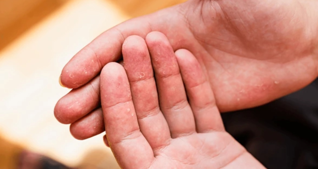
Initial Manifestations
Keratolysis exfoliativa typically begins with the appearance of small, superficial air-filled blisters on the palmar surface of the hands and fingers. These initial lesions are generally asymptomatic and may go unnoticed until they rupture, leading to characteristic skin peeling.
Seasonal Patterns and Triggers
The condition demonstrates a notable seasonal pattern, with approximately 50% of cases occurring during warmer months, particularly in individuals with palmar hyperhidrosis. This seasonal variation supports the role of heat and humidity in disease pathogenesis.
Disease Progression
The progression of keratolysis exfoliativa follows a predictable pattern. Initial white macules evolve into “corneal layer vesicles” or “dry vesicles” that subsequently break in the center. As peeling occurs, collarettes of paper-thin scales form at the edges of lesions, which clinically resemble superficial flaccid vesicles that have recently ruptured.
Distribution and Affected Areas
While primarily affecting the palms, exfoliative keratolysis can occasionally involve the soles of the feet and, less commonly, the dorsal surfaces of hands and feet. The peeling typically occurs in circular or oval shapes, leaving behind tender, erythematous areas that may be susceptible to secondary infections.
Characteristic Features
Unlike other dermatological conditions, keratolysis exfoliativa is characterized by a lack of inflammation and minimal associated symptoms. Eventually, most of the palmar surface may become affected as older lesions coalesce and new lesions appear.
Differential Diagnosis and Associated Conditions
Primary Differential Diagnoses
Accurate diagnosis of keratolysis exfoliativa requires careful differentiation from several similar conditions. The most important differential diagnoses include dyshidrotic eczema, contact dermatitis, tinea manuum, epidermolysis bullosa simplex, and acral peeling skin syndrome.
Distinguishing Features
Unlike these conditions, keratolysis exfoliativa lacks the inflammatory component typically seen in eczematous conditions and does not respond to topical corticosteroids. Dyshidrotic eczema presents with deeper vesicles that contain fluid and are associated with significant itching and inflammation.
Additional Diagnostic Considerations
Contact dermatitis typically shows a clear relationship to allergen exposure and demonstrates positive patch test results. Tinea infections can be ruled out through negative potassium hydroxide (KOH) preparations and fungal cultures. The lack of genetic mutations and the superficial nature of the peeling help distinguish keratolysis exfoliativa from epidermolysis bullosa simplex.
Associated Conditions
Exfoliative keratolysis may be associated with atopic dermatitis in some individuals, though this relationship remains unclear. Primary hyperhidrosis is frequently observed in affected patients, with associated excessive sweating reported in 58-61% of cases.
Treatment Approaches and Management Strategies
Conservative Management Principles
The management of keratolysis exfoliativa focuses primarily on symptomatic relief and prevention of exacerbating factors. Since the condition is self-limited and typically resolves spontaneously, treatment aims to expedite healing and prevent complications.
Irritant Avoidance
The cornerstone of keratolysis exfoliativa treatment involves identification and avoidance of irritants, including soaps, detergents, solvents, and excessive water exposure. This preventive approach forms the foundation of long-term management.
Topical Therapies
Topical emollients form the foundation of therapeutic intervention. Effective moisturizers should contain ingredients that both hydrate and promote gentle exfoliation of dead skin cells. Recommended formulations include those containing urea, lactic acid, or silicone compounds.
Specialized Topical Treatments
Urea-containing creams at concentrations of 20-40% provide both moisturizing and keratolytic effects. Ammonium lactate 12% cream and salicylic acid 6% cream offer similar benefits in promoting skin turnover and hydration.
Advanced Treatment Options
For more resistant cases of keratolysis exfoliativa, healthcare providers may consider additional therapeutic modalities. Photochemotherapy (PUVA treatment) has shown efficacy in severe cases. Oral acitretin, a systemic retinoid, has demonstrated marked dose-response improvement in extensive disease cases that failed to respond to standard topical therapy.
Corticosteroid Limitations
Topical corticosteroids generally provide limited benefit since keratolysis exfoliativa lacks significant inflammatory components. When prescribed, potent corticosteroids such as betamethasone dipropionate 0.05% or clobetasol 0.05% should be used for no more than two weeks.
Extreme Cases and Complications
Severe Disease Presentation
While keratolysis exfoliativa in extreme cases is relatively uncommon, it can present significant challenges for both patients and clinicians. Extensive disease may involve large portions of the palmar and plantar surfaces, leading to widespread desquamation and potential functional impairment.
Complications in Severe Cases
In such cases, patients may experience painful fissuring of the affected areas, making daily activities difficult and potentially leading to secondary bacterial infections. Complications in severe cases may include chronic skin sensitivity, increased susceptibility to contact irritants, and impaired barrier function.
Advanced Treatment for Extreme Cases
Keratolysis exfoliativa in extreme cases may require more aggressive therapeutic intervention. The use of systemic acitretin has proven particularly valuable in these scenarios, with patients showing marked improvement in skin texture and reduction in peeling episodes.
Drug-Induced Cases
Drug-induced keratolysis exfoliativa represents another form of extreme presentation. A documented case involving ranolazine, a cardiac medication, demonstrated how certain pharmaceuticals can trigger keratolysis exfoliativa-like eruptions.
Epidemiological Patterns and Risk Factors
Demographic Distribution
The epidemiology of keratolysis exfoliativa reveals interesting demographic patterns. Studies indicate that the condition affects both sexes, with a slight female predominance (female to male ratio of approximately 1.2:1).
Age Distribution
The peak incidence occurs in young adults, particularly in the 21-30 age group, accounting for 25.3% of all cases. A secondary peak is observed in the 11-20 and 31-40 age groups.
Occupational Risk Factors
Occupational factors play a significant role in disease prevalence. Housewives represent the largest affected demographic group at 30%, likely due to frequent exposure to water, soaps, and household chemicals. Manual laborers and individuals in occupations requiring frequent hand washing also show increased incidence rates.
Environmental Influences
Environmental factors contribute significantly, with higher prevalence during warm, humid months when sweating and friction are more pronounced. The seasonal variation of exfoliative keratolysis supports the role of heat and humidity in disease pathogenesis.
Geographic and Cultural Factors
Geographic and cultural factors may influence disease presentation. Saltwater fishermen have been reported to experience higher rates of the condition, though whether this relates to saltwater exposure or bacterial contact from fish handling remains unclear.
Prevention Strategies and Lifestyle Modifications
Primary Prevention Measures
Preventing keratolysis exfoliativa recurrences requires a comprehensive approach addressing known triggering factors. Primary prevention focuses on minimizing exposure to irritants and maintaining optimal skin barrier function.
Hand Hygiene Modifications
Hand hygiene practices require modification in susceptible individuals. Rather than using hot water, which can strip natural skin oils, lukewarm water should be used for hand washing. The duration of water exposure should be minimized, and hands should be thoroughly dried after washing.
Moisturizing Protocols
Moisturizing routines form a crucial component of prevention strategies. Regular application of barrier creams containing silicone compounds, petrolatum, or mineral oil can protect against irritant exposure. These skin protectants should be applied particularly after hand washing and before activities involving potential irritant contact.
Climate Control Strategies
Climate control measures can reduce seasonal exacerbations. In humid environments or during summer months, efforts to maintain dry conditions and minimize excessive sweating prove beneficial. Air conditioning, fans, and moisture-wicking clothing can help reduce the heat and humidity that trigger episodes.
Prognosis and Long-term Outlook
Overall Prognosis
The prognosis for keratolysis exfoliativa is generally excellent, with most cases following a benign, self-limiting course. Episodes typically last from several days to weeks, followed by complete skin regeneration in affected areas.
Recurrence Patterns
The condition tends to be recurrent, with new episodes appearing every few weeks or months, particularly during triggering seasons or with irritant exposure. Long-term complications are rare in typical cases of keratolysis exfoliativa.
Age-Related Improvements
The natural history of exfoliative keratolysis often shows improvement with age. Many patients report decreased frequency and severity of episodes over time, possibly due to changes in hormonal status, occupational exposures, or lifestyle modifications.
Patient Education Importance
Patient education plays a crucial role in optimizing long-term outcomes. Understanding the benign nature of the condition helps reduce anxiety and prevents inappropriate treatments that might worsen symptoms.
Advanced Therapeutic Considerations
Emerging Treatment Modalities
Recent advances in understanding keratolysis exfoliativa pathophysiology have opened new therapeutic avenues. The recognition of premature corneodesmolysis as the primary mechanism has led to the investigation of treatments targeting corneodesmosomal stability.
Phototherapy Applications
Phototherapy represents an emerging treatment modality for recalcitrant cases. Hand and foot PUVA therapy has shown promise in reducing episode frequency and severity. The mechanism involves photoactivation of psoralen compounds, which may stabilize corneodesmosomal connections and normalize skin turnover.
Systemic Treatment Options
Systemic retinoids, particularly acitretin, have demonstrated efficacy in keratolysis exfoliativa in extreme cases. The treatment targets abnormal corneocyte desquamation and has shown marked dose-response relationships.
Novel Topical Formulations
Research into novel topical formulations continues to evolve. Barrier repair creams containing ceramides and other lipid components may help restore normal skin architecture. Additionally, anti-inflammatory agents that don’t rely on corticosteroid mechanisms are being investigated for their potential to reduce tissue damage associated with chronic peeling episodes.
Conclusion
Keratolysis exfoliativa represents a fascinating example of how subtle disruptions in normal skin physiology can lead to distinctive clinical presentations. While the condition remains incompletely understood from an etiological perspective, recent advances in histopathological and molecular research have provided valuable insights into its pathogenesis. The management of keratolysis exfoliativa continues to evolve from purely symptomatic approaches to more targeted interventions addressing underlying pathophysiology. While most cases remain mild and self-limiting, the availability of advanced treatments such as phototherapy and systemic retinoids provides options for severe cases that might otherwise cause significant functional impairment. Patient education and preventive strategies remain cornerstones of effective management, with emphasis on irritant avoidance and appropriate moisturizing regimens. The generally excellent prognosis and benign nature of the condition should provide reassurance to affected individuals, while the availability of effective treatments ensures that even challenging cases can be successfully managed with appropriate dermatological care.