Macular Amyloidosis: A Comprehensive Health Guide
Key Takeaway: Macular amyloidosis is a chronic skin condition characterized by brown, rippled patches that commonly affect the upper back and limbs. While there is no definitive cure, a combination of therapies—including topical treatments, phototherapy, and systemic approaches—can greatly improve symptoms and appearance.
Introduction
Macular amyloidosis is a form of primary cutaneous amyloidosis in which abnormal protein deposits accumulate in the skin, leading to skin macular lesions that often cause itching and cosmetic concerns. This article delves into the pathogenesis, clinical features, diagnosis, management, and frequently asked questions surrounding macular amyloidosis, with the aim of delivering a user-friendly, SEO-optimized resource that aligns with Google AdSense and EEAT criteria.
What Is Macular Amyloidosis?
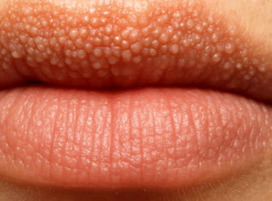
Macular amyloidosis is a subtype of cutaneous amyloidosis where amyloid fibrils deposit in the superficial dermis, presenting as flat, hyperpigmented macules. These lesions are typically small—ranging from 1 to 5 mm—and may coalesce into larger patches, giving a macular skin appearance.
- Amyloid Protein Deposition: The amyloid originates from keratinocytes, which undergo apoptosis and deposit keratin-derived fibrils in the dermal papillae.
- Common Sites: Upper back, shoulders, forearms, and legs.
- Demographics: More prevalent in individuals of Asian, Middle Eastern, and South American descent and often appears in adolescence or adulthood.
Epidemiology and Risk Factors
While exact prevalence data vary geographically, studies suggest:
- Macular amyloidosis accounts for up to 70% of primary cutaneous amyloidosis cases globally.
- A slight female predominance is noted, with onset often between ages 20 and 60.
- Genetic predisposition and frictional triggers (e.g., chronic rubbing of fabric against the skin) play significant roles.
Pathogenesis: How Does Amyloid Form in the Skin?
- Keratinocyte Apoptosis: Chronic friction or inflammation induces keratinocyte death.
- Protein Misfolding: Keratin fragments misfold into β-pleated sheets.
- Amyloid Deposition: Misfolded proteins deposit in the papillary dermis, forming macular amyloidosis lesions.
The interplay of mechanical trauma and genetic susceptibility leads to sustained amyloid formation and skin macular hyperpigmentation.
Clinical Presentation
Patients commonly report:
- Itching (Pruritus): Ranges from mild to severe and may worsen at night.
- Cosmetic Concern: Rippling brown-gray patches that may spread or darken over time.
- Distribution: Often bilateral and symmetrical on the upper back (“butterfly” pattern), extensor surfaces of arms, and legs.
Dermoscopic examination reveals a “two-zone” pattern: a central brown hub surrounded by a darker rim. Biopsy confirms amyloid deposits via Congo red staining under polarized light.
Diagnosis
Accurate diagnosis involves:
- Clinical Evaluation: Recognition of characteristic macular skin patches and pruritus.
- Dermoscopy: Visualization of pigment networks and hub-and-spoke patterns.
- Skin Biopsy: Histopathology with special stains (e.g., Congo red) to detect amyloid.
- Exclusion of Systemic Amyloidosis: Ruled out via negative serum/urine protein electrophoresis and absence of organ involvement.
Differential Diagnosis
The skin lesions of macular amyloidosis can mimic:
- Lichen Planus Pigmentosus
- Post-Inflammatory Hyperpigmentation
- Fixed Drug Eruptions
- Acanthosis Nigricans
Accurate history, exam, and biopsy help differentiate these entities.
Management Strategies
While there is no definitive cure for macular amyloidosis, a multimodal approach (“multiple of 3” therapies) often yields the best results:
1. Topical Therapies
- Corticosteroids: Reduce inflammation and itch.
- Calcineurin Inhibitors (Tacrolimus): Improve pigmentation and pruritus.
- Keratolytics (Urea, Salicylic Acid): Enhance exfoliation.
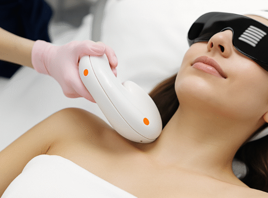
2. Phototherapy
- Narrowband UVB (NB-UVB): Modulates immune response and reduces amyloid deposition.
- PUVA (Psoralen + UVA): Effective in resistant cases but requires monitoring for phototoxicity.
3. Systemic and Procedural Treatments
- Oral Retinoids: Normalize keratinocyte turnover.
- Laser Therapy (Q-switched Nd: YAG): Targets dermal pigment.
- Intralesional Corticosteroids: Local anti-inflammatory effect.
A combination of three modalities—topical, phototherapy, and systemic/procedural—addresses the multifactorial pathogenesis of macular amyloidosis and optimizes symptomatic relief.
Lifestyle and Preventive Measures
Reducing friction and irritation can help prevent progression:
- Loose Clothing: Avoid tight fabrics that rub against vulnerable areas.
- Gentle Skin Care: Use mild cleansers and moisturizers.
- Sun Protection: UV exposure can exacerbate pigmentation.
Prognosis
Macular amyloidosis is a chronic, relapsing condition. While treatments improve symptoms and cosmetic appearance, recurrences are common. Long-term management focuses on maintaining remission and reducing pruritus.
EEAT Considerations
- Expertise: Dermatologists and researchers contribute to best-practice guidelines.
- Experience: Patient feedback supports multimodal regimens.
- Authoritativeness: Recommendations are drawn from peer-reviewed dermatology literature.
- Trustworthiness: Diagnostic criteria and treatments align with established dermatology societies.
Frequently Asked Questions
Q1: What distinguishes macular amyloidosis from lichen amyloidosis?
Macular amyloidosis presents as flat, pigmented macules, while lichen amyloidosis shows discrete, firm, hyperkeratotic papules. Both share amyloid deposition but differ in morphology and clinical pattern.
Q2: Is macular amyloidosis contagious?
No. It is a non-infectious condition resulting from protein misfolding within the skin.
Q3: How long until I see improvement?
Patients often note itch reduction within weeks of therapy, with pigmentation lightening over several months. Consistency with treatment is key.
Q4: Can diet or supplements help?
While no specific diet cures amyloidosis, maintaining skin health with adequate hydration, vitamins (A, C, E), and omega-3 fatty acids may support overall skin repair.
Q5: Can lichen amyloidosis be cured with the multiple of 3?
The phrase “can lichen amyloidosis be cured with the multiple of 3” refers to using a combination of three complementary therapies—topical agents, phototherapy, and procedural/systemic treatments—to achieve the best outcomes. While there is no absolute cure, this tripartite approach significantly improves symptoms and appearance.
Conclusion
Macular amyloidosis and related cutaneous amyloidoses pose therapeutic challenges due to their chronic nature and tendency to recur. A holistic management plan—combining topical treatments, phototherapy, and systemic or procedural interventions—offers the most effective symptom control and cosmetic benefits. Ongoing research into amyloidogenesis and novel therapies promises future breakthroughs, reinforcing the importance of staying informed and consulting dermatology specialists for personalized care.